Treatment Summary
Patient Information
- Age: 37
- Gender: male
Diagnosis
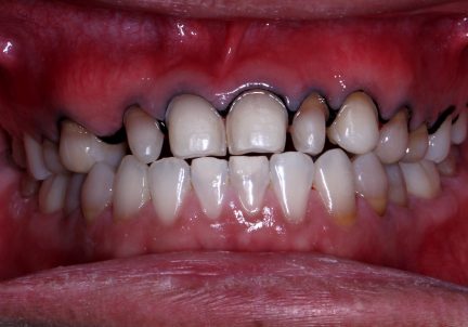
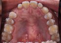
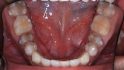
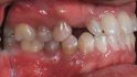
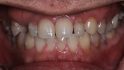
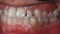
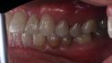
- Post eruptive color changes of dental hard tissues on 1.1 (UR1), 1.2 (UR2), 1.3 (UR3), 1.4 (UR4), 1.5 (UR5), 2.1 (UL1), 2.2 (UL2), 2.3 (UL3), 2.4 (UL4), 2.5 (UL5)
- Cracked tooth on 1.1 (UR1), 2.1 (UL1)
Total Treatment Visits
- 9
Treatment planning
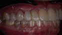
Appointment 1: After pre-restorative aligner treatment the patient had a whitening treatment done. It was performed to correct Tetracycline-associated staining of the teeth
Appointment 2: After 2 weeks, a direct mock-up was placed on the unprepped teeth. The patient was sent back to try the fit and occlusion
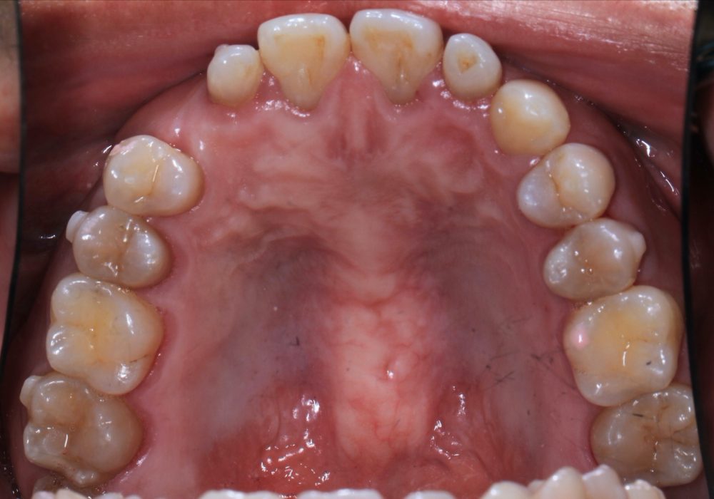
Appointment 3: After 2 weeks teeth preparation on #15, #14, #12, #11, #21, #22, #23, #24, and #25 was done and scans were taken and sent to the laboratory
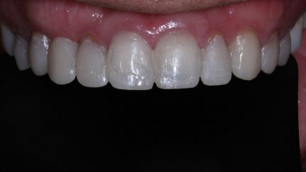
Appointment 4: Cementation of the veneers on teeth #15, #14, #12, #11, #21 , #22, #23, #24 and #25
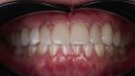
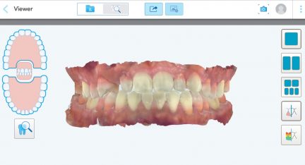
Appointment 5: Follow-up to check the fit of the veneers. An iTero scan was taken to plan the implant crown
Appointment 6: Implant surgery on #13 was completed
Appointment 7: After 3 months, digital impression of #13 for manufacturing the crown.
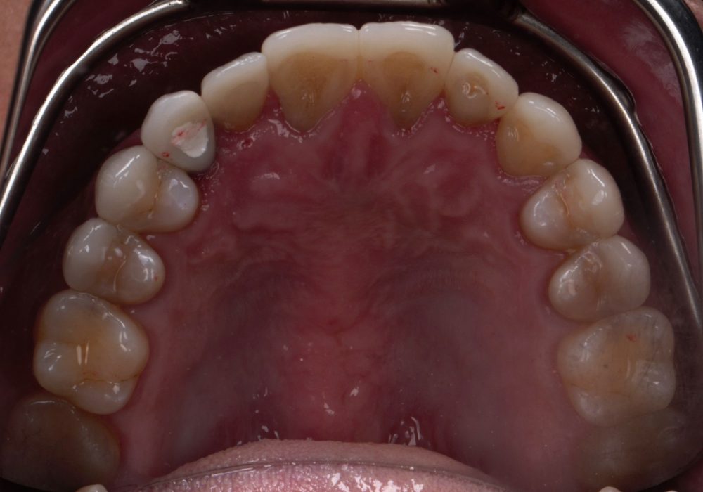
Appointment 8: Cementation of single implant crown on #13
Appointment 9: Follow-up appointment
Comments
Dental history: Pt. had missing #13 and anterior crossbite. Pre-restorative aligner treatment consisted of 16 upper aligners and 38 lower aligners. Additional 8 aligners for both upper and lower jaws were ordered for minor corrections.
Chairside milling was done for the final veneers
The treatment was seamless, predictable, and, accurate with the effective use of digital technology


 316
316  310
310 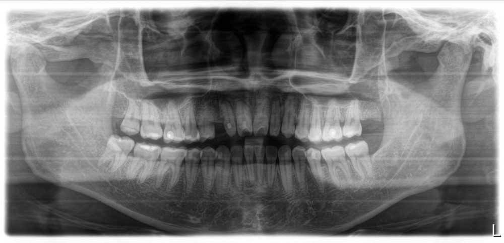 310
310 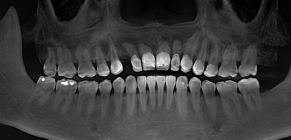 304
304