Treatment Summary
Patient Information
- Age: 60
- Gender: male
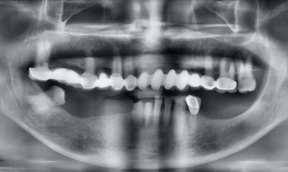
Diagnosis
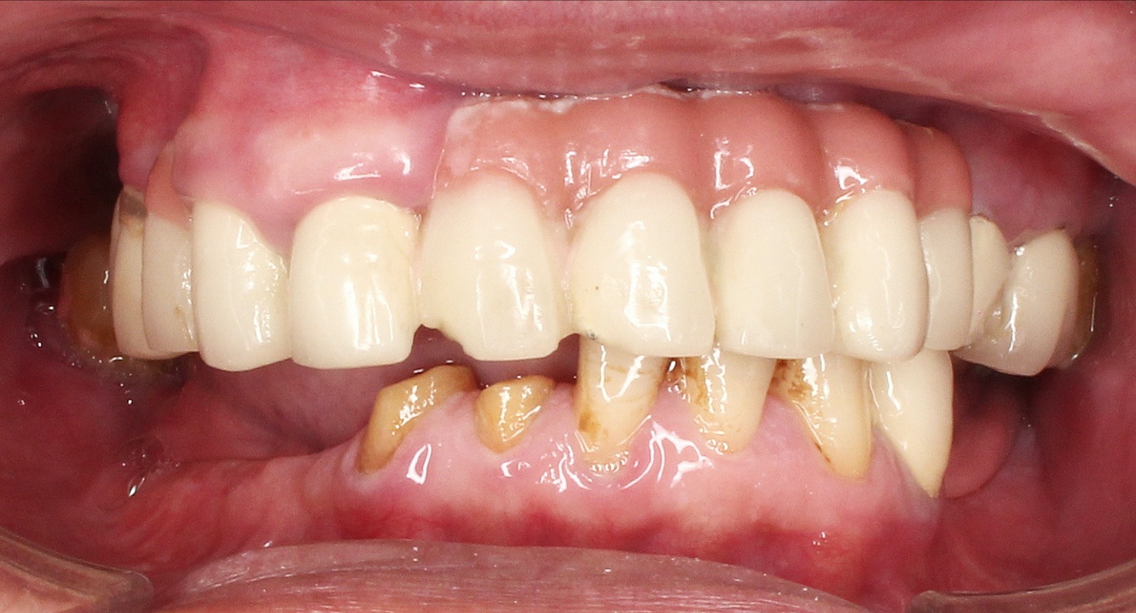
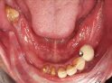
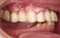
- Abrasion on 3.1 (LL1), 3.2 (LL2), 3.3 (LL3)
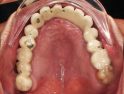
- Horizontal alveolar bone loss on 1.1 (UR1), 1.4 (UR4), 2.1 (UL1), 2.2 (UL2), 2.3 (UL3), 2.4 (UL4), 3.5 (LL5), 3.6 (LL6), 3.7 (LL7), 3.8 (LL8)
- Complete loss of tooth on 1.1 (UR1), 1.4 (UR4), 1.6 (UR6), 1.7 (UR7), 2.1 (UL1), 2.2 (UL2), 2.3 (UL3), 2.4 (UL4), 2.8 (UL8), 4.1 (LR1), 4.4 (LR4), 4.5 (LR5), 4.6 (LR6), 4.7 (LR7), 3.5 (LL5), 3.6 (LL6), 3.7 (LL7), 3.8 (LL8)
- Partial loss of tooth on 4.2 (LR2), 4.3 (LR3)
- Unsatisfactory restoration on 1.1 (UR1), 1.2 (UR2), 1.3 (UR3), 1.4 (UR4), 1.5 (UR5), 1.6 (UR6), 1.7 (UR7), 1.8 (UR8), 2.1 (UL1), 2.2 (UL2), 2.3 (UL3), 2.4 (UL4), 2.5 (UL5), 2.6 (UL6), 3.4 (LL4)
Total Treatment Visits
- 11
Treatment planning
Step 1:
- Clinical examination, intra and extra-oral photographs followed by CBCT, and treatment plan was devised
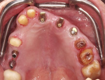
- Removal of an old bridge and dental abutments planned for teeth #15, #13, #12, #25, and #26
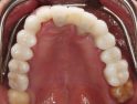
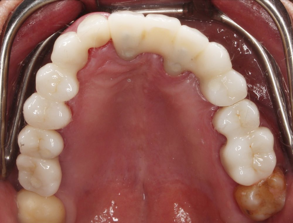
- Implants were placed in the position of #16, #17, #11, #22, #24
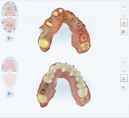
- iTero scan of abutments was taken for immediate PMMA bridge fabrication
Step 2:
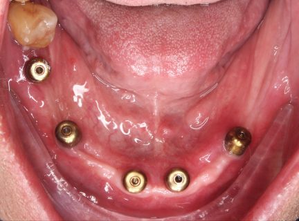
-Extraction of #31, #32, #33, #34, #42, #43
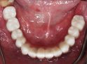
- 5 implants were placed on the lower jaw
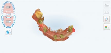
- Scans were taken for immediate loading of a temporary lower denture.
Step 3: Immediate loading of the lower denture within a week from implant placement
Step 4: Post 6 months of healing, scan the upper and lower for the final prosthesis
Step 5: Try in of upper bridges and lower titanium CAD/CAM framework, and check for centric relation
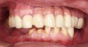
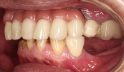
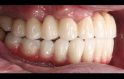
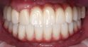
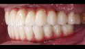
Step 6: Final prosthesis try-in and cementation, adjusting the occlusion and polishing
Comments
Applying digital impression protocol with iTero scanner for final prostheses:
- For the upper arch: scan dental abutments and scan bodies for fabricating zirconia bridges
- For the lower arch: scan multiunit scan bodies for fabricating fixed prosthesis
- Scan of PMMA bridges, dentures, and occlusion to replicate the provisional prosthesis in the final design.


 310
310 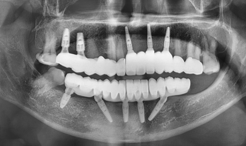 304
304