Treatment Summary
Patient Information
- Age: 36
- Gender: female
Diagnosis
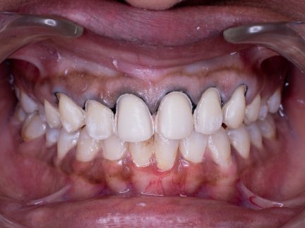
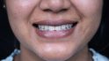
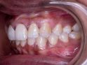
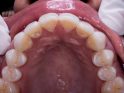
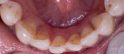
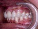
- Post eruptive color changes of dental hard tissues on 1.1 (UR1), 1.2 (UR2), 1.3 (UR3), 1.4 (UR4), 2.1 (UL1), 2.2 (UL2), 2.3 (UL3), 2.4 (UL4)
Total Treatment Visits
- 5
Treatment planning
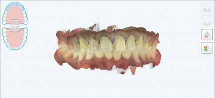
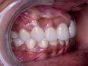
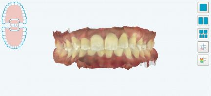
Step 1: Consultation using the iTero intraoral scanner, followed by intraoral photographs.
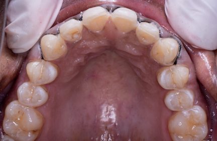
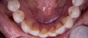
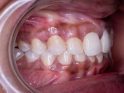
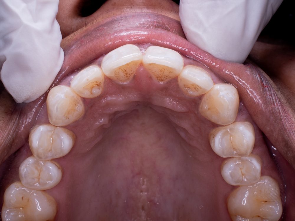
Step 2: Teeth preparation was done on #14, #13, #12, #11, #21, #22, #23 and #24.
Step 3: Gingival retraction was done using 000 braided cords soaked in hemostal.
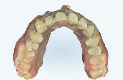
Step 4: iTero scanner was used to scan prepped teeth in high-resolution.
Step 5: Digital mock-up was created and approved.
Step 6: e.max veneers were fabricated.
Step 7: Intaglio surface of the veneers was etched with hydrofluoric acid and then the primer was applied.
Step 8: Prepped teeth were etched with 37% phosphoric acid and a bonding agent was applied and cured.
Step 9: Veneer cementation was done, tack cured, the excess was removed and then the interproximal space was cleared using a dental floss.
Step 10: Complete curing was done.
Yorumlar
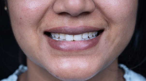
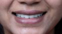
History: The patient had a chief complaint of discolored teeth, white patches and irregularly shaped teeth
Treatment plan: Indirect veneers were planned on the upper arch from #14 to #24


 398
398